Understanding of biomineralization processes
Biomineralization is defined as the ability of living organism to build mineralized tissues, most of the time to protect or support soft tissues. Biological mineralized tissues are often hybrid materials where biogenic minerals are intimately associated to an extra cellular matrix (ECM), composed of biomacromolecules directing the 3D repartition of biominerals. The main biominerals characteristics (composition, phase, size, morphology, surface properties…) are biologically or physico-chemically controlled. However, the underlying processes are still largely unknown. Hence, we use solid state NMR to investigated biogenic minerals and mineralized tissues to understand the ultrastructure and the formation mechanisms at to the atomic scale.
I. Ultrastructure of mineralized tissues
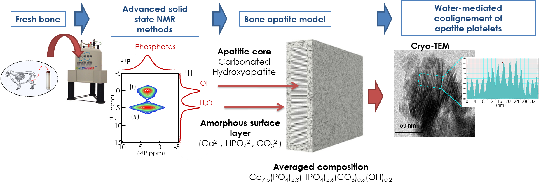
The hierarchical organization of biominerals is controlled at several length scales by the ECM and the corresponding template macromolecules (protein or polysaccharides). Biomineral surface is exposed to the ECM components and then influences the mechanical, metabolic or remodelling properties of the mineralized tissue. As a striking example, bone is composed of a calcium phosphate phase associated to fibrillar collagen. The mineral phase is close to hydroxyapatite (Ca10(PO4)6(OH2) in terms of composition but differs significantly as it is known to be “Ca deficient”, “poorly crystalized” and “highly substituted”. Bone mineral surface was largely unknown and, by studying fresh bone sample, we evidenced its core-layer organization where the surface layer can be considered as an independent mineral domain, both on a structural and chemical point-of-view. If the core is found as a carbonate-substituted hydroxyapatite phase, surface domain is close to an amorphous calcium phosphate (ACP) composed of divalent ions (Ca2+, HPO42-, CO32-). This extensive study of structural bone mineral description allowed us to update the chemical composition of bone apatite. Further, working on native hydration preserved samples and using complementary techniques such as WAXS and Cryo-TEM led us to evidence strongly adsorbed water molecules considered as a “hydration shell” surrounding bone mineral crystals and directing the alignment of apatite platelets along their c-axis direction through the strong interaction with the ACP layer.

Funding: ANR NanoShap (2009–2012), Labex Matisse (2013-2016), PHC Ulysse (2019)
PhD Students: Yan Wang (2009-2012), Stanislas Von Euw (2011-2014)
Main Collaborations: N. Nassif (LCMCP), G. Pehau-Arnaudet (Institut Pasteur), C. Drouet (CIRIMAT Toulouse)
Articles:
Y. Wang, S. Von Euw et al. Nat. Mater. 2013
S. Von Euw et al. Acta Biomater. 2017
S. Von Euw et al. Scientific Reports 2019
We are using similar approach for calcium carbonate-based mineralized tissues such as nacre or urchin spines. Nacre is composed of aragonite crystals stacked in-between sheets of chitin. Once again, organo-mineral interface determines mechanical properties of that material. As an example, fracture resistance is 3000 times higher compared to single crystal aragonite. We have shown through advanced solid-state NMR study of European abalone Haliotis tuberculata that, unlike bone apatite, nacre aragonite surface is not totally amorphous and the organo-mineral interface is not mediated by a layer of water molecules.

Funding: Labex Matisse (2015–2018), ARED Région Bretagne (2015-2018), PEPS CNRS (2017)
PhD Students: Widad Ajili (2015-2018)
Main Collaborations: N. Nassif (LCMCP), S. Auzoux-Bordenave (BOREA MNHN), N. Menguy (IMPMC), Y. Politi (B CUBE Dresden)
Articles:
M. Albéric et al. Crystal Growth & Design 2018
W. Ajili et al. J. Phys. Chem. C 2020
II. Dynamic Nuclear Polarization for the comprehension of biomineralization processes
DNP is a recent technique allowing a huge gain in sensitivity for the NMR signal. This technique has been adapted both to liquid-state NMR, i.e. dissolution DNP, and to solid state NMR, i.e. MAS DNP. In the latter case, the use of exogenous polarization agent enables to reveal surfaces of materials. We recently demonstrated that MAS DNP can bring atomic-scale detailed informations on the organo-mineral interface both in bone and nacre. 31P-13C REDOR experiments boosted by DNP are achieved within hours instead of weeks! The involvement of citrate functions in bone is revealed together with their adsorption mode. Similarly, the role of bicarbonates at the organo-mineral interface in nacre is evidenced thanks to MAS DNP.

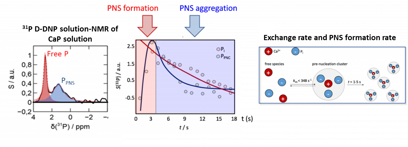
NMR is a robust method to determine the structure of molecules in solution. However its poor intrinsic sensitivity is limiting the access to fast chemical processes. Using dissolution DNP (D-DNP) monitoring, we probed, in collaboration with Dennis Kurzbach and his team (Vienna University), fast interaction kinetics such as those underlying the formation of pre-nucleation species (PNS) that develop within milliseconds when calcium and phosphate ions meet in solution and that precede non-classical solid-liquid phase separation. Such PNS are described as (meta)stable ionic clusters and their aggregation leads the nucleation of calcium phosphate (or carbonates) phases. In case of oversaturated CaP solutions, PNS exhibit a very limited lifetime as these transient precursors form and disappear on milliseconds time-scale. Nonetheless, D-DNP enables the characterization of such transient PNS on structural (size) and dynamical (exchange and formation kinetic) point-of-view.

Funding: CNRS-NTU Excellence Science (2019)
Main Collaborations: Anne Lesage group (Lyon), Dennis Kurzbach group (Vienna)
PhD Students: Tristan Georges (2019-2022)
Articles:
T. Azaïs et al. Solid State Nucl. Magn. Reson.2019
E. Weber et al. Anal. Chem. 2020
III. Synthesis of biomimetic minerals
In order to fully understand biomineralization processes it is of paramount importance to model biogenic minerals formation through biomimetic synthesis mimicking physiological conditions which includes the control of the ionic concentrations, the macromolecules liquid-crystalline phase or the presence of acidic mineralizing proteins. Nucleation, formation and phase transformation/crystallization can be studied in situ, by inducing the phase transformation within the NMR rotor, or ex situ, by sampling at relevant time points. Several parameters directing the crystallization of calcium phosphate or carbonate phases have been evaluated including pH, level of hydration, physical confinement within macromolecular matrices… Further, complementary techniques such as WAXS and Cryo-TEM enable the comprehension of apatite platelets self-assembly.
Funding: Labex Matisse (2015–2018), ANR NanoShap (2009–2012), Labex Matisse (2013-2016), PHC Ulysse (2019)
Collaboration: G. Costentin & J-M. Kraft (LRS), S. Von Euw (Trinity College Dublin), G. Pehau-Arnaudet (Institut Pasteur), G. Renaudin (ICCF Clermont-Ferrand)
PhD Students: Marc Robin (2015-2018), Yan Wang (2009-2012), Stanislas Von Euw (2011-2014)
Articles:
Y. Wang et al. Mater. Horiz. 2014
S. Von Euw et al. Geoscience 2018
M. Robin et al. CrystEngComm 2020
S. Von Euw et al. J. Am. Chem. Soc. 2020
